As we all know the Digestive system is one of the important topics of biology. Here we have briefly discussed the human digestive system, the parts involved in the digestive system, and the workings of the human digestive system. The digestive system processes food, breaking it down into nutrients for absorption and elimination of waste through organs like the stomach, small and large intestines, and liver.
Human Digestive System
The Human digestive system is responsible for breaking down food into nutrients that can be absorbed and used by the body. It includes organs such as the mouth, esophagus, stomach, small intestine, large intestine, and more, each organ plays a specific role in digestion and nutrient absorption. The process of digestion involves both mechanical and chemical actions to convert food into smaller molecules that can be transported through the bloodstream for energy and growth.

Human Digestion
Digestion is the third step of Holozoic nutrition and it is a breakdown of large complex insoluble molecules into smaller, simple, and soluble molecules which are easy to absorb. These simple and smaller molecules are ultimately taken into the blood and then into the cells to be utilized for metabolic needs by the body. Digestion takes place with the help of many digestive enzymes. Digestion is of two types: Intracellular digestion and Extracellular digestion
Intracellular Digestion
Intracellular digestion refers to the digestion process that occurs within the confines of a cell. In this process, the cells break down the food particles within them. Intracellular digestion takes place in a specialized compartment called Lysosome, where enzymes break the food molecules into smaller nutrients that the cell can absorb and use. It is commonly found in single-cell organisms. This type of digestion is found in Protozoans and in some multicellular animals like Hydra, Echinoderms, etc.
Extracellular Digestion
The digestion which takes place outside the cell in a narrow cavity called the digestive tract is known as Extracellular digestion. It is common in most multicellular organisms and animals including some insects, fungi, and certain aquatic organisms. In Extracellular digestion, the digestive enzymes are secreted into the external environment, where the breaking down of complex food molecules into simpler ones takes place.
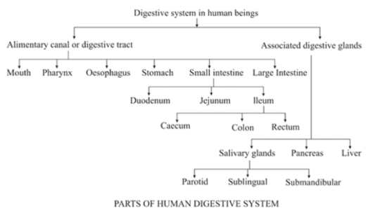
Parts of the Human Digestive System
The human digestive system consists of two parts: the Alimentary Canal and Associated Glands. Each part has its own important function. Both the alimentary canal and associated glands work together to break down food, absorb nutrients, and eliminate waste from the body. The Alimentary Canal and the Associated gland are briefly discussed below.
| Parts of the Human Digestive System | |
| Parts | Description |
| Alimentary Canal | The alimentary canal, or digestive tract is a muscular tube that processes food from the mouth to the anus, aiding digestion and nutrient absorption. |
| Associated Glands | Associated glands, like the salivary, liver, and pancreas, secrete enzymes and substances into the altimetry canal to aid digestion and nutrient breakdown. |
Alimentary Canal
The alimentary canal is also known as the digestive tract or gastrointestinal tract. The alimentary canal is a long muscular tube (about 8-9 meters in length) of varying diameter. It extends from the mouth to the anus. The digestion and absorption of food are functions performed by the alimentary canal. It includes the following structures:
- Mouth and Buccal Cavity
- Esophagus
- Stomach
- Small Intestine
- Large Intestine
Mouth and Buccal Cavity
The mouth is the opening or cavity in the lower part of the human face. It is also known as the Buccal cavity or Oral cavity, as the food enters the body through it. The mouth is bounded by two soft movable lips. Within the buccal cavity, there exists a muscular tongue and teeth positioned across two sets of jaws.
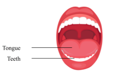
(a) Tongue: The tongue is a thick muscular-sensory organ present on the floor of the buccal cavity. It has various taste buds which help to detect the taste. The surface is covered by a mucous membrane. The upper lateral surface is covered with various types of Papillae such as Fungiform Papillae, Filiform Papillae, and Vallate Papillae.
- Filiform Papillae: are conical projections distributed in a parallel row over the anterior two-thirds of the tongue. They have a whitish appearance and lack taste buds.
- Fungiform Papillae: Mushroom-like projections are scattered amidst the Filiform Papillae, with a higher concentration towards the tongue’s tip, and the majority of these projections house taste buds.
- Vallate Papillae: are arranged in the form of an inverted ‘V’ on the posterior lateral surface of the tongue and contain taste buds.
Taste buds are those cells that are present in the tongue. They are the small cavities that open to the surface through Gustatory pores. The taste buds in different parts of the tongue respond to particular modalities of taste such as sweet, sour, bitter, and salty.
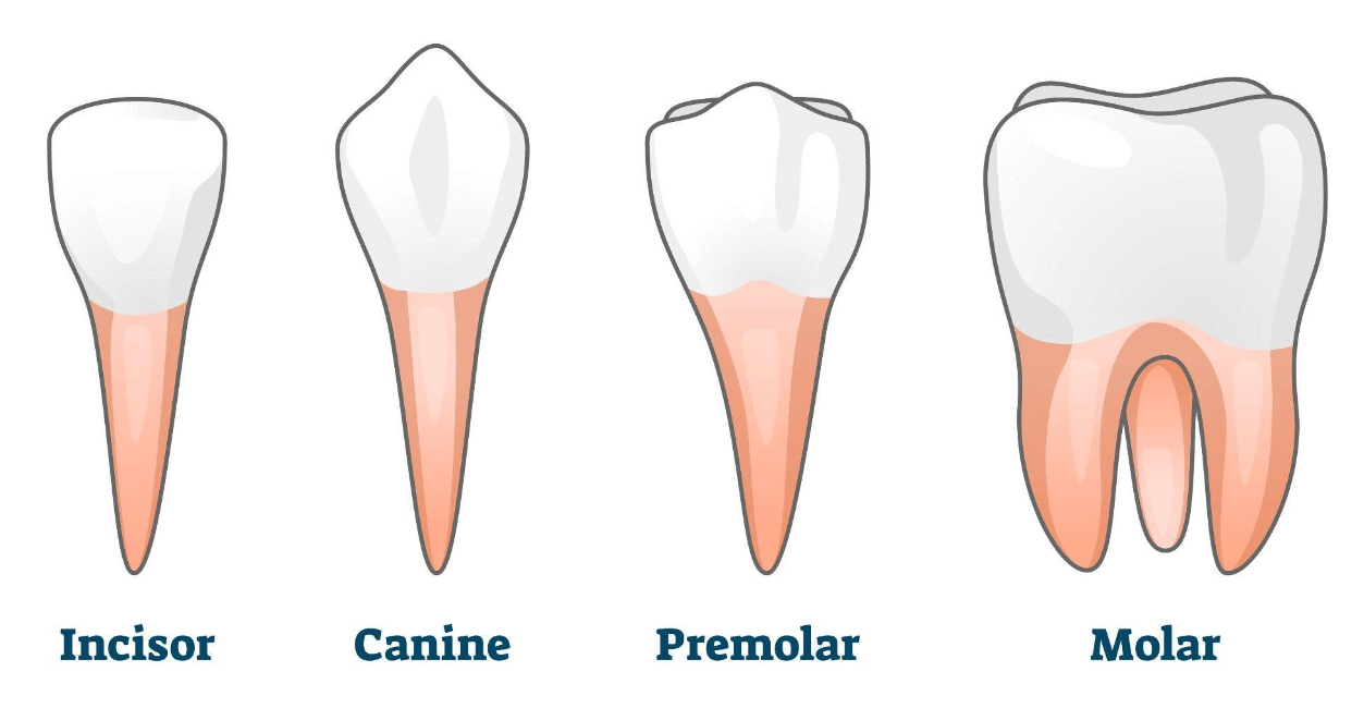
(b) Teeth: An adult human has 16 teeth in each jaw, embedded in Jaw- sockets. They appear twice in a lifetime. The first are the milk teeth and they are later replaced by the permanent teeth in adults which last for years. There are four different types of teeth: incisors, canines, premolars, and molars. The dental formula of an adult human is 2123 i.e., in each half of the jaw, from middle to backward. There are two incisors, one canine, two premolars, and three molars.
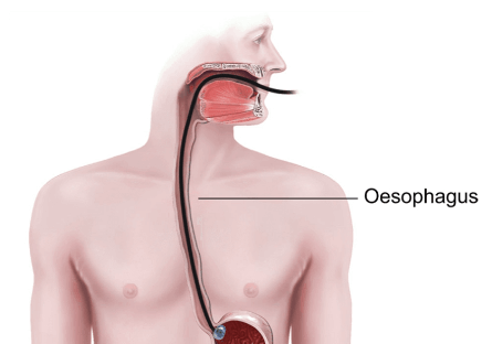
Oesophagus
The esophagus is 22-25 cm long, narrow, muscular, and tubular structure. The esophagus runs downward through the neck behind the trachea and opens in the stomach in the abdomen. The esophagus plays a crucial role in transporting food and liquids from the mouth to the stomach for digestion. The swallowed food is moved to the stomach by the peristaltic movement of esophageal muscles present in the Esophagus.
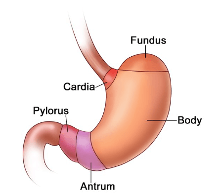
Stomach
The stomach is a large, muscular, and somewhat J- J-shaped sac. The stomach occupies the left side of the upper part of the abdominal cavity. The upper part of the stomach is called the Cardiac, the middle dome-shaped part is the Body, and the distal part is the Pyloric. The stomach has two ends: The cardiac end is the end where the stomach is attached to the Oesophagus and the pyloric end is where the stomach is attached to the Duodenum. There are numerous glands in the stomach which are called gastric glands.
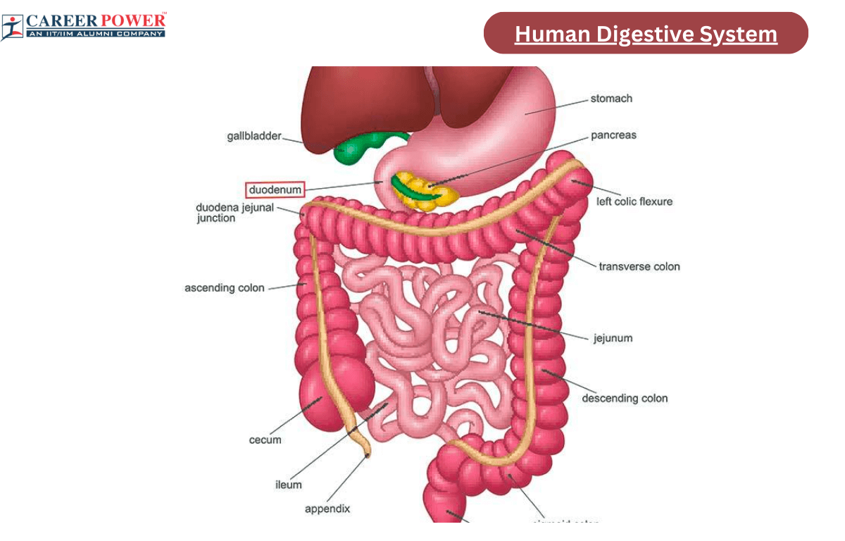
Small Intestine
The small intestine is the longest part of the Alimentary Canal, about 6 meters in length. The small intestine occupies the central and lower part of the abdominal cavity. It is divided into three regions. which are: The Duodenum, Jejunum, and Ileum.
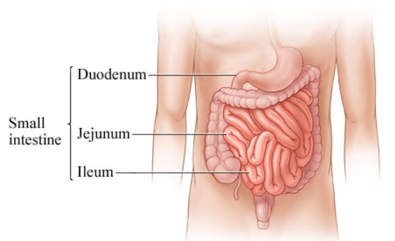
- Duodenum: The first part of the small intestine is called Duodenum. It looks somewhat like the alphabet “C”. The ducts which convey the pancreatic juice as well as the duct which carries Bile open in the duodenum.
- Jejunum: The duodenum opens into Jejunum, which is about 2.5 meters long.
- Ileum: The ileum is the longest part of the small intestine and it opens in the caecum in the lower part of the abdominal cavity. Both Ileum and Jejunum are highly coiled structures. The inner mucosa of the small intestine is raised into millions of minute finger-like projections, called villi.
Large Intestine
The Ileum opens into the large intestine. The large intestine is comparatively shorter in length but wider in diameter when compared to the small intestine. The large Intestine does not possess any villi. The large intestine is divided into the following three regions that are: Caecum, Colon, and Rectum.
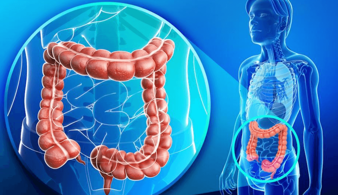
- Caecum: The caecum is a small, pouch-like structure that ends into a tubular part like the vermiform appendix. Both the caecum and the vermiform appendix are vestigial in function and are not involved in cellulose digestion.
- Colon: The colon is a long sacculated structure that is differentiated into the Ascending colon, extending up to the liver on the right side; the Transverse colon, which crosses the abdomen cavity below the pancreas; the Descending colon, running downwards on the left side; Pelvic colon, which is “S” shaped, continuous into the rectum.
- Rectum: The rectum is slightly dilated and about 13 cm long. It opens outside by the anus and is guarded by two anal sphincter muscles.
Associated Glands
The Associated gland generally refers to glands that are closely connected or related to a specific organ or system in the body. These glands often work together to regulate various functions of the body. Associated glands help in the process of digestion by releasing several enzymes. The Associated Glands are divided into five types which are: Salivary Glands, Gastric Glands, Liver, Pancreas, and Intestinal Glands.
- Salivary Glands: Historically salivary glands are racemose glands and they look like a cluster of grapes. Each gland consists of a large number of acini. The function of the salivary gland is to secrete salivary juice, the salivary gland secretion is rich in salivary amylase, a starch-splitting enzyme that helps in the breakdown of carbohydrates. There are three types of salivary glands: parotic, submaxillary, and sublingual.
- Parotic: These are the largest of all the salivary glands and are located near the ears. The parotic glands are responsible for the production of saliva which leads to digestion and oral hygiene.
- Submaxillary: These generally refer to the submandibular glands, which a salivary glands located beneath the lower jaw. Like other salivary glands, it also helps in the production of Saliva.
- Sublingual: Sublingual refers to the administration of medication or substances under the tongue, where they are absorbed directly into the bloodstream with the help of the mucous membrane.
- Gastric Glands: These gastric glands are present in the stomach. Gastric juices secreted by the gastric glands play a great role in the process of digestion. Gastric glands are of three types Fundic, Pyloric, and Cardiac Glands.
- Fundic glands are present in the body and fundus region of the stomach and secret hydrochloric acid (HCL), pepsinogen, and soluble mucin. The secretion is highly acidic.
- Pyloric glands are found in the pyloric region of the stomach. The secretion is rich in mucin but does not contain hydrochloric acid and is slightly alkaline. these glands also secrete a hormone called gastrin.
- Cardiac glands are found in the cardiac region of the stomach and secrete mucin and a small amount of pepsinogen.
- Liver: The liver is the largest gland of the body, weighing about 2.5 kg. The liver is located in the right upper part of the abdomen just below the diaphragm. On the lower surface of the right liver lobe, a thin-walled pear-shaped sac is present which is called Gall bladder. The liver acts as an exocrine gland for the secretion of bile. It also acts as a storehouse for several substances like glucose, lipids, vitamins, and iron. It helps in the removal of unwanted matters from the blood.
- Pancreas: The pancreas is a gland that is partly exocrine and partly endocrine, but the main gland consists of a head, neck, body, and tail. The head of the pancreas fits in the “C” shaped curve of the duodenum. Its body extends to the left and its tail touches the spleen. The endocrine part of the pancreas is in the form of numerous rounded collections of cells that are embedded within the exocrine part. These collections of cells are called the Islet of Langerhans.
- Intestinal glands: The intestinal glands are present in the mucosa of the small intestine and are divided into two types: Crypts of Lieberkuhn and Brunner’s Gland
(a) Crypts of Lieberkuhn: These are simple tubular glands, opening into the lumen of the small intestine. It contains some mucus-secreting Goblet cells, the juice produced by these glands is known as Succus Entericus.
(b) Brunner’s Glands: These are compound tubular glands that are found only in the submucosa of the duodenum and open in the space in between the villi. These glands secrete alkaline mucus-rich juice.
Working of the Human Digestive System
The human digestive system is a complex series of organs and processes that help in the breakdown of food into nutrients that can be absorbed and used by the human body. The digestion of carbohydrates starts in the mouth itself with the help of saliva. The working of the human digestive system involves several steps. Here is the step-by-step working of the human digestive system:
Step 1: Ingestion- Ingestion is the first step or we can say that the digestion process begins with Ingestion. It is the intake of food through the mouth.
Step 2: Mouth- Mechanical digestion starts in the mouth as the teeth break down the food into smaller pieces. Saliva contains enzymes like salivary amylase, which starts the chemical digestion of carbohydrates.
Step 3: Pharynx and Esophagus- The chewed food forms a bolus and is pushed into the Pharynx, which leads to the esophagus. The muscular contractions (Parastalsis) move the bolus down the esophagus to the stomach.
Step 4: Stomach- The stomach churns and mixes food with gastric juices, which include hydrochloric acid and pepsin. These gastric juices begin the breaking down of proteins. The final mixture is called Chyme.
Step 5: Small Intestine- Chyme enters the small intestine, where most digestion and absorption take place. Bile from the liver and enzymes from the pancreas helps in the breakdown of fats, proteins, and carbohydrates in the small intestine.
Step 6: Large Intestine- Water and electrolytes are reabsorbed from the undigested food, and this leads to the formation of feces.
Step 7: Rectum- Feces are stored until elimination.
Step 8: Anus- The elimination of feces takes place through the anus by the process called Defecation.


 50 Vegetables Name for Kids in English a...
 Food Chain: Definition, Types, Examples,...
 Human Respiratory System: Definition, Di...