Respiration is how our bodies get energy from food. It happens in two main steps: breathing and cellular respiration. When we breathe, we take in oxygen and release carbon dioxide. In our cells, oxygen combines with food molecules to produce energy, water, and carbon dioxide. This energy helps our body function. Carbon dioxide is then removed when we breathe out. Here we have briefly described the human respiratory system, its parts, and the process below in the article.
What is the Respiratory System?
The respiratory system is a biological system that allows living organisms to exchange gases, primarily oxygen and carbon dioxide, with their environment. In humans, this system includes the nose, nasal passages, pharynx, larynx, trachea, bronchi, and lungs. When we breathe in, oxygen is taken into the lungs, where it is transferred to the bloodstream and carried to cells throughout the body. At the same time, carbon dioxide, a waste product of cellular metabolism, is removed from the blood and exhaled from the body when we breathe out. This exchange of gases is essential for the body’s cells to function properly and is a fundamental process for sustaining life.
Human Respiratory System
The human respiratory system is like our body’s breathing engine. It includes the lungs and a network of tubes called bronchi and bronchioles. When we breathe in, air travels down these tubes into the lungs, where oxygen from the air gets into our bloodstream, and carbon dioxide, a waste product, goes out. This oxygen fuels our body and helps it work, while the waste carbon dioxide is released when carbon dioxide is released when we breathe out.
In addition to the lungs and tube, the respiratory system also has tiny air sacs called Alveoli, where the exchange of oxygen and carbon dioxide takes place. When we breathe, the Diaphragm, a Muscle below our Lungs, contracts and relaxes, allowing air to enter and leave. This continuous process provides our body with the oxygen it needs to function and removes harmful carbon dioxide, ensuring our cells stay healthy and our body can perform daily activities.
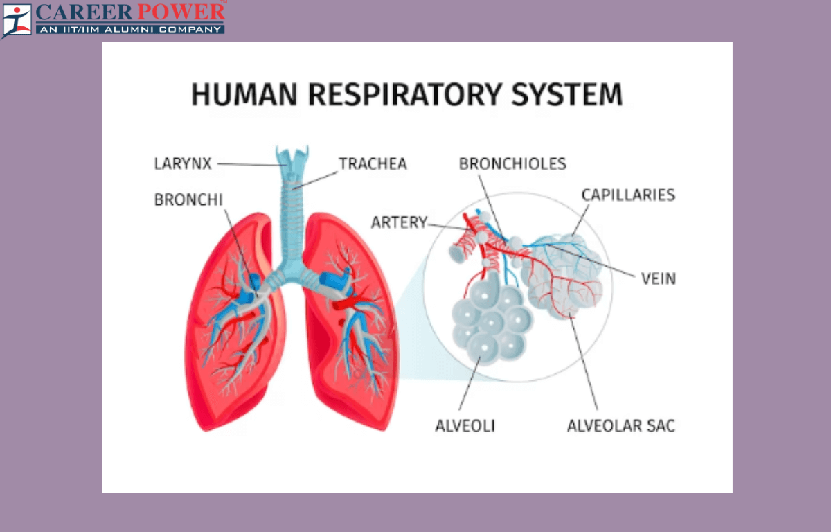
Diagram of a Human Respiratory System
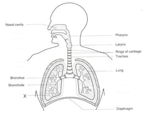
The diagram of the human respiratory system illustrates the organs and structures responsible for breathing. It typically includes the nasal cavity, trachea, bronchi, and lungs. Inhaling air through the nose, travels down the trachea, branching into the bronchi, and finally reaching the lungs. There, oxygen is exchanged for carbon dioxide in tiny air sacs called alveoli, enabling the body to oxygenate blood and remove waste gases.
Parts of the Human Respiratory System
The human respiratory system is a complex network of organs and tissues that helps us breathe. It consists of several parts that work together to facilitate the process of breathing and exchange of gases. Here we have discussed some of the main components of the respiratory system. These components work together to ensure the exchange of oxygen and carbon dioxide, supporting the body’s oxygen needs and removing waste gases.
| Different Parts of Respiratory System | |
| Components of the Respiratory System | Description |
| Nose and Nasal Cavity | The nose and nasal cavity are filters, that moisten and warm incoming air. |
| Pharynx | The pharynx works as a passage for both air and food, connecting the nasal cavity, mouth, and larynx. |
| Larynx | The larynx contains vocal cords; involved in speech and airway protection. |
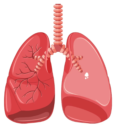
| Trachea (Windpipe) | The trachea carries air from the larynx to the lungs, which is reinforced with cartilage rings. |
| Bronchi | Bronchi are two branches from the trachea, leading to each lung. |
| Bronchioles | Bronchioles are smaller branches of bronchi within the lungs. |
| Alveoli | Alveoli are tiny sacs where gas exchange occurs; oxygen in, carbon dioxide out. |
| Lungs | Lungs are the main respiratory organs, responsible for oxygenating blood. |
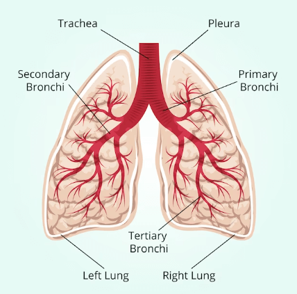
| Pleura | Pleura are the membranes surrounding the lungs and lining the chest cavity, creating a pleural cavity. |
| Diaphragm | The diaphragm is a dome-shaped muscle below the lungs, that aids in breathing by expanding and contracting. |
Nose and Nasal Cavity
The nose and nasal cavity are very important as air is taken in through the nose, where it is filtered, moistened, and warmed by tiny hair-like structures called cilia and mucous membranes in the nasal cavity.
Pharynx
The pharynx is also known as the throat, the pharynx serves as a passage for both air and food. The pharynx connects the nasal cavity and the mouth to the larynx.
Larynx
The larynx is commonly known as the voice box. The larynx contains the vocal cords and is involved in speech production. It also acts as a value to close the airway during swallowing, preventing food and liquids from entering the trachea.
Trachea
The trachea is also known as the windpipe. The trachea is a tube-like structure that carries air from the larynx to the lungs. Its walls are reinforced with cartilage rings to keep it open and prevent collapsing.
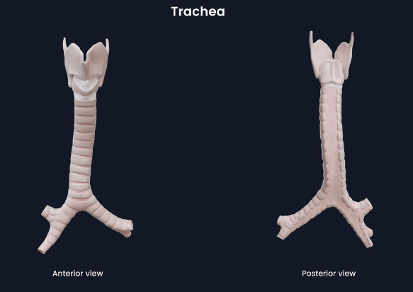
Bronchi
The trachea divides into two bronchi, where each bronchi leads to each lung. These bronchi further divide into smaller tubes called bronchioles.
Bronchioles
Bronchioles are smaller branches of the bronchi within the lungs. They continue to divide into even smaller tubes and eventually lead to tiny air sacs called alveoli.
Alveoli
Alveoli are small, grape-like clusters of air sacs at the end of the bronchioles. They are the site of the gas exchange, where oxygen from the air enters the bloodstream, and carbon dioxide, a waste product of metabolism, exits the bloodstream to be exhaled.
Lungs
The lungs are the main organs of the respiratory system and are located in the chest cavity. They are responsible for inhaling oxygen and exhaling carbon dioxide. The right lung has three lobes, while the left lung has two lobes to accommodate the space occupied by the heart.
Pleura
The pleura are thin membranes that surround each lung and line the chest cavity. They create a lubricated space (pleural cavity) between them, allowing the lungs to expand and contract smoothly during breathing.
Diaphragm
The diaphragm is a large, dome-shaped muscle located at the base of the lungs and plays a crucial role in breathing. When it contracts and flattens, the volume in the chest cavity increases, causing inhalation. When it relaxes, the volume decreases, leading to exhalation.
Working of the Human Respiratory System
Here we have discussed a step-by-step explanation of how the human respiratory system works. This continuous process of inhalation, gas exchange, and exhalation ensures that the body receives the oxygen it needs and eliminates carbon dioxide, a waste product of cellular metabolism.
Step 1: Breathing In (Inhalation)
- Nose and Mouth: Air is taken in through the nose and/or mouth.
- Nasal Passage: Air passes through the nasal passages, where it is filtered, humidified, and warmed.
- Pharynx: The air then moves into the pharynx, a passage shared with the Digestive System.
- Larynx: The air travels through the larynx, where vocal cords are located.
- Trachea: From the larynx, the air enters the trachea, a large tube reinforced with cartilage rings.
Step 2: Conducting Airways
- Bronchi: The trachea divides into two bronchi (one for each lung), which further divide into smaller bronchi and bronchioles.
- Bronchioles: These smaller tubes lead to tiny air sacs called alveoli.
Step 3: Gas Exchange (Alveoli)
- Alveoli: At the end of bronchioles, there are millions of tiny air sacs called alveoli. This is where the actual gas exchange occurs.
- Capillaries: Alveoli are surrounded by tiny blood vessels called capillaries. Oxygen from the inhaled air diffuses across the alveolar membrane into the blood, binding with hemoglobin in red blood cells.
- Carbon Dioxide (CO2) Exchange: At the same time, carbon dioxide, a waste product produced by cells, diffuses from the blood into the alveoli to be exhaled.
Step 4: Breathing Out (Exhalation)
- Diaphragm and Intercostal Muscles: Exhalation is a passive process where the diaphragm and intercostal muscles relax, decreasing the volume of the chest cavity.
- Air Expulsion: The decrease in volume increases the air pressure in the lungs, forcing air out of the respiratory system through the same pathway it came in.
Step 5: Gas Transport
- Blood Circulation: Oxygen-rich blood is carried away from the lungs by pulmonary veins and is pumped by the heart to various parts of the body.
- Delivery of Oxygen: In the body’s tissues, oxygen is released from hemoglobin and used for cellular respiration, a process where cells produce energy from nutrients.
- Collection of CO2: Deoxygenated blood, now rich in CO2, returns to the heart and is pumped back to the lungs for exhalation.
Functions of the Respiratory System
The respiratory system performs several vital functions in the human body. These functions are essential for maintaining the body’s oxygen supply, removing waste gases, and supporting overall physiological processes.
- Breathing (Ventilation): The main function is to facilitate the exchange of oxygen and carbon dioxide between the body and the environment. When you breathe in (inhale), oxygen from the air enters your lungs. When you breathe out (exhale), carbon dioxide, a waste product of metabolism, is expelled from your body.
- Gas Exchange: In the lungs, oxygen from the inhaled air diffuses into the bloodstream, where it binds to hemoglobin in red blood cells. At the same time, carbon dioxide, which is carried in the bloodstream, diffuses from the blood into the lungs and is expelled when you exhale.
- Regulation of Blood pH: By controlling the levels of carbon dioxide in the body, the respiratory system helps regulate the blood’s pH (acid-base balance). This balance is crucial for normal function and metabolism.
- Voice Production: The respiratory system, particularly the larynx, enables humans to produce sounds and speak.
- Protection Against Microorganisms: The respiratory system has mucus-producing cells and tiny hair-like structures called cilia, which help trap and remove dust, microbes, and other particles from the respiratory tract. This helps protect the lungs from infections.
- Sense of Smell: The nasal cavity, a part of the respiratory system, contains olfactory receptors that enable the sense of smell.
Some Important Questions Regarding the Human Respiratory System
Below, we have discussed a few important questions from the chapter Human Respiratory System that might be asked in the exams.
Q1. What are the important respiratory system parts in humans?
Answer: The most important human respiratory system parts include the Nose, larynx, pharynx, trachea, bronchi, and lungs.
Q2. What are the main functions of the Human respiratory system?
Answer: The main functions of the respiratory system include- inhalation and exhalation of gases, exchange of gases between the bloodstream and lungs, the gaseous exchange between the bloodstream and body tissues, olfaction, and vibration of vocal cords.
Q3. What are the steps involved in the process of the human respiratory system?
Answer: The steps involved in the process of the human respiratory system include: Breathing In (Inhalation), Conducting Airways, Gas Exchange (Alveoli), Breathing Out (Exhalation), Gas Transport.


 50 Vegetables Name for Kids in English a...
 Food Chain: Definition, Types, Examples,...
 Animal Kingdom: Classifications, Example...