The main transport system in Human Beings is the Circulatory System. The circulatory system is counted as one of the important topics in Biology. In the Circulatory System, the blood carries oxygen, digested food, and other chemicals like hormones and enzymes to all the parts of the body. The Human Circulatory System consists of the Heart, Veins, Arteries, Capillaries, and Blood.
The Heart is the vital organ that plays a great role in receiving and pumping blood, while the Arteries, Capillaries, and Veins act like a pipe or tube through which the blood flows in the body. All the Arteries, Veins, and Capillaries are together known as Blood Vessels, which are responsible for the proper functioning of the heart. In Human beings, various substances are transported through two liquids which are “blood” and “lymph”. Here we have provided a brief overview of the human heart in detail.
The Human Heart
The Heart is the most important part of the circulatory system, as it receives and pumps blood to every part of the body. The Heart makes a “Lub-Dub” sound during rhythmic contraction and relaxation. The heart operates continuously throughout a person’s lifetime and is the most industrious muscle within the human body. It provides oxygen and nutrients to various cells in the body while assisting in the removal of waste products.
Almost all living organisms possess a heart that pumps blood throughout their bodies. Even birds, reptiles, and amphibians possess a heart-like pumping organ, but it does not function the same way in every organism, as the heart of birds only has three chambers, and fishes have two chambers in their hearts.
Structure of the Human Heart
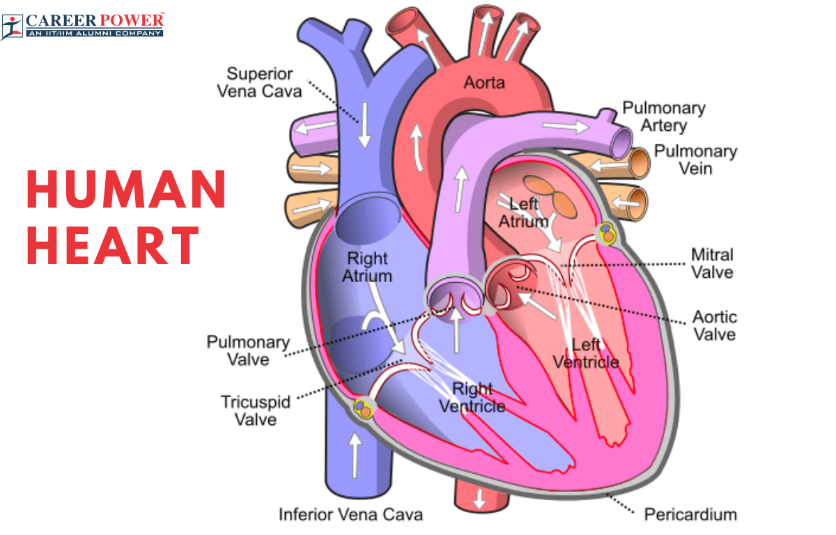
The Heart is the main pumping organ of the body, it regulates the proper flow of blood. It is located slightly left of the center of the chest and is protected by the ribcage. The heart is shaped like an upside-down pear, kind of like a cone. It is made of special muscles called cardiac muscles. The size of the heart is about the same as a clenched fist. Most arteries carry oxygen-rich blood, but the pulmonary artery is an exception as it carries oxygen-poor blood. Most veins carry oxygen-poor blood, but the pulmonary vein is different because it carries oxygen-rich blood.
Chambers of the Heart
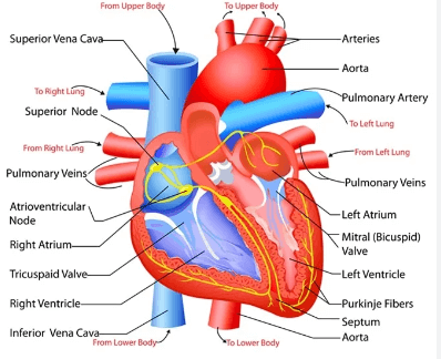
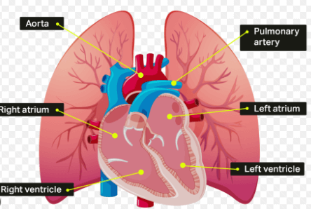
The Human Heart has four compartments known as “Chambers” inside it, which are named the Right atrium, Left atrium, Right ventricle, and Left ventricle.
| Chambers of the Human Heart | ||
| Chambers | Location | Function |
| Left Atrium | The left atrium is located at the upper left side of the heart. | Receives oxygenated blood from the lungs via pulmonary veins. |
| Left Ventricle | The left ventricle is located at the lower left side of the heart | Pumps oxygenated blood to the rest of the body through the aorta. |
| Right Atrium | The right atrium is located at the upper right side of the heart. | Receives deoxygenated blood from the body through the superior and inferior vena cava. |
| Right Ventricle | The right ventricle is located at the lower right side of the heart. | Pumps deoxygenated blood to the lungs for oxygenation through pulmonary arteries. |
These chambers are together known as:
- Atria
- Ventricles
Atria
The upper two chambers of the heart are called Atria which are singly called Right Atrium and Left Atrium. These atria are the fundamental components of the Circulatory System. They are often referred to as the receiving chambers of the heart. Situated at the top of the heart, the atria are designed to receive blood from two different sources: the right atrium receives deoxygenated blood from the body through the superior and inferior vena cava while the left atrium receives oxygenated blood from the lungs through the pulmonary veins.
Ventricles
The lower two chambers are the Ventricles which are singly known as the Right Ventricle And Left Ventricle. These ventricles are often referred to as the primary pumping chambers of the heart, which carry blood to the entire body and lungs. The right ventricle pumps the deoxygenated blood to the lungs through the pulmonary artery and the left ventricle pumps oxygenated blood to the rest of the body through the aorta. The aorta is the largest and the main artery in the Human Body. The Ventricles are separated by the interventricular Septum, which is a muscular wall that prevents the mixing of oxygenated and deoxygenated blood.
Heart Valves
The heart consists of special structures within the heart that ensure the one-way flow of blood. The Human Heart consists of four valves that help in the proper functioning and flow of blood through the chambers. These walls open and close with each heartbeat; the opening, and closing process of the heart valves prevents blood from flowing backward. The four valves of the heart are named as Aortic valve, Mitral valve, Pulmonary valve (or pulmonic valve), and Tricuspid valve. The functioning and origin of the heart valves have been discussed below.
| Valves Present in the Human Heart |
||
| Valves | Location | Function |
| Aortic valve | Between the left ventricle and the aorta | Controls blood flow from the left ventricle into the aorta and prevents backflow into the ventricle. |
| Mitral valve | Between the left atrium and left ventricle | Allows blood to flow from the left atrium into the left ventricle and prevents backflow. |
| Pulmonary valve | Between the right ventricle and the pulmonary artery | Controls blood flow from the right ventricle into the pulmonary artery and prevents backflow into the ventricle. |
| Tricuspid valve | Between the right atrium and right ventricle | Allows blood to flow from the right atrium into the right ventricle and prevents backflow. |
Tricuspid valves
The tricuspid valves are one of the heart’s four valves. These tricuspid valves are located between the right atrium and right ventricle. These tricuspid valves have leaflets that open to allow blood flow through the atrium to the ventricle and close to prevent backflow of the blood.
Pulmonary valves
Pulmonary valves are located at the exit of the right ventricle and it is the entrance to the Pulmonary artery (which carries deoxygenated blood from the heart to the lungs). The pulmonary valves open to allow blood flow from the right ventricle into the pulmonary artery and then closes, so that there is no backflow of the blood.
Mitral or Bicuspid valve
The bicuspid valves are located between the left ventricle and the left atrium. They play a crucial role in preventing the backflow of blood from the left ventricle. Normally the heart valves have three leaflets, but in some individuals, the bicuspid valves have two leaflets. This condition is present from the birth itself. It is very important for individuals with bicuspid valves to maintain a healthy lifestyle.
Aortic valves
The aortic valves are located at the exit of the left ventricle and it is the entrance to the Aorta. These Aortic valves prevent the backflow of blood back into the left ventricle from the Aorta.
Functions of the Human Heart
The heart plays many functions in the body. Pumping of blood is the primary function, the heart plays many secondary functions. This multiple functioning of the heart makes it a crucial organ for overall health. The other functions are mentioned below.
- Pumping of blood: The heart serves as a muscular pump that contracts rhythmically. The heart’s main function is to pump blood throughout the body. The right side of the heart receives deoxygenated blood from the body while the left side receives oxygenated blood from the lung and then pumps it into the rest of the body.
- Regulation of Blood Pressure: The heart helps in the regulation of blood pressure, by regulating the pumping process. Adequate blood pressure is essential for ensuring that blood reaches all parts of the body.
- Removal of unwanted substances: The heart helps in the removal of metabolic waste and carbon dioxide from the body through the bloodstream.
- Transportation of Hormones: The heart also plays a role in the transportation of hormones to various tissues and organs.
- Releases Stress Hormones: When a person is stressed, the heart releases a stress hormone (which is the same as adrenaline), this stress hormone prepares the body for a “fight or flight” response.
Working of Human Heart
The continuous pumping of blood is known as the Cardiac Cycle, and this cardiac cycle is controlled by Electrical impulses. These electrical impulses are generated by the natural pacemaker of the heart which is known as the Sinoatrial (SA) node.
Step 1: The blood enters the heart through two large veins, the superior vena cava (from the upper body) and the inferior vena cava (from the lower body). This deoxygenated blood enters the right atrium.
Step 2: When the right atrium contracts, it pushes the blood to the right ventricle through the tricuspid valve.
Step 3: The right ventricle contracts and the deoxygenated blood passes through the pulmonary valve with the help of the pulmonary artery.
Step 4: The blood is passed to the lungs through the pulmonary artery, in the lungs the blood picks up oxygen and releases carbon dioxide. Now the blood becomes oxygenated.
Step 5: This oxygenated blood then comes back to the heart’s left atrium through the pulmonary veins.
Step 6: The left atrium contracts and pushes the oxygenated blood to the left ventricle through the mitral valve.
Step 7: The left ventricle pumps oxygen-rich blood through the aortic valve with the help of the aorta. The left ventricle is the strongest chamber of the human heart.
Step 8: Aorta is the largest artery of the human body and it distributes oxygenated blood to all the parts of the body by a complex network of blood vessels.
Heart Beats
One complete contraction and relaxation of the heart is called a heartbeat. The heart rate of a healthy human is 60-100 beats per minute (bpm). The heart rate can vary based on facts such as age, fitness level, and overall health. We can even feel our heartbeats if we place our hand on the chest just above the heart region, while a doctor listens to our heartbeat with the help of an apparatus called a Stethoscope. The heart beats faster during and after heavy workouts because the body needs more energy under these conditions.
Heart Pulse Rate
The blood is forced into arteries, every time the heart beats. This blood expands the arteries. The expansion of an artery each time the blood is forced into it is the Pulse. The pulse rate of a person is equal to the number of heartbeats per minute. The pulse rate is essentially used to measure the heart rate and is an indicator of overall cardiovascular health. We can feel the pulse at the wrist, and neck just by slightly pressing the artery with our fingertips, as the arteries are close to the surface of the skin.
Blood Pressure
Blood pressure is the pressure at which blood is pumped around the body by the heart. Blood pressure is expressed in two forms “Systolic pressure” and “Diastolic pressure”. The maximum pressure at which the blood leaves the heart during contraction is called Systolic pressure. The minimum pressure at which the blood leaves the heart through the main artery Aorta is called Diastolic pressure. If a person is suffering from high blood pressure then it is called hypertension, while low blood pressure is called Hypotension. The blood pressure is measured in millimeters of mercury (mm Hg). The normal systolic and diastolic values are:
- Systolic Pressure: 120 mm Hg
- Diastolic Pressure: 80 mm Hg


 50 Vegetables Name for Kids in English a...
 Food Chain: Definition, Types, Examples,...
 Human Respiratory System: Definition, Di...