As we all know that the human body consists of five sense organs- Eyes, Nose, Ears, Tongue, and Skin. The Eyes let us see things around us. The Nose lets us smell different scents. The Ears help us hear sounds. The Tongue allows us to taste flavors. The Skin lets us feel things like touch and temperature. Each of these organs helps us understand and interact with the world in different ways. All these sense organs are interlinked with each other and are controlled by the Human Brain. We have discussed more about the human eye, its parts, and its functions in the article below.
Human Eye
The Human eye works on the reflection of light through a natural convex lens made of transparent living material and helps us to see things around us. Our eyes, which are very important and sensitive tools for seeing, are considered a special gift. If we didn’t have eyes, we could somewhat figure out objects by their taste, smell, and sound, but we wouldn’t be able to tell their color.
The eyes are like a natural camera, their lens system forms images on the light-sensitive screen called a retina. There are many different complex and sensitive parts of the human eye. In our article, we have discussed the Parts of the Eye and Their Functioning.
Parts of the Eye
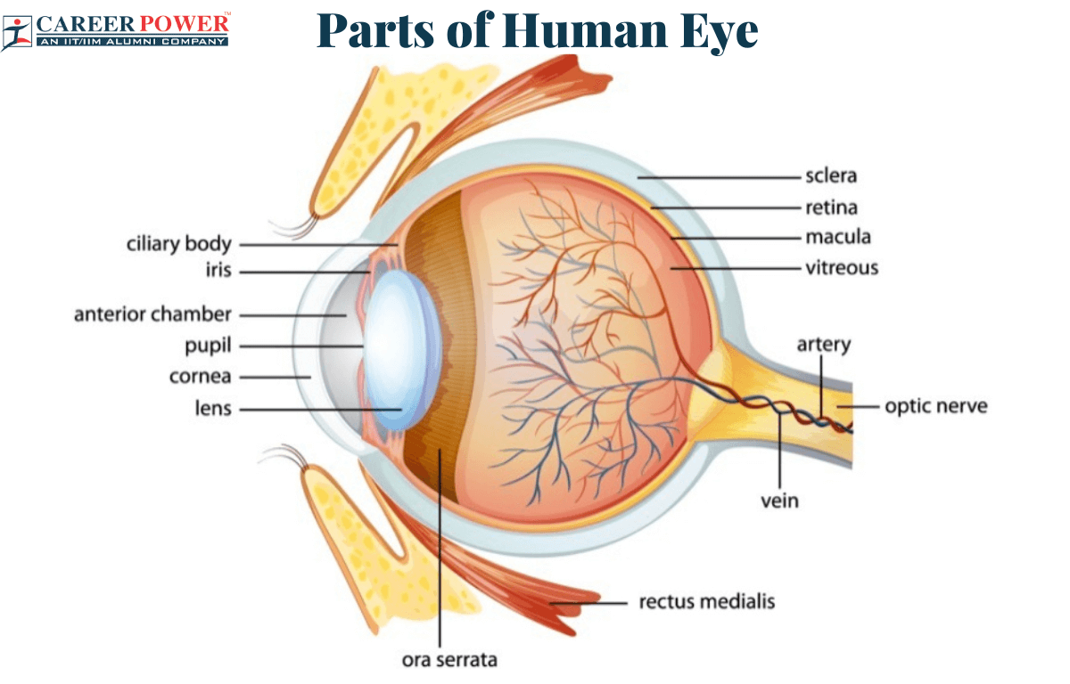
The human eye consists of 9 parts and each part plays an important role and works together to allow us to see and process visual information. The eyeball is spherical in shape having a diameter of about 2.5 cm. For a clear image, an object should be placed in the correct position. The minimum distance at which an object must be placed so that the normal eye can see it clearly without any strain is called the least distance of distinct vision. The distinct vision for a normal human eye is 25 cm.
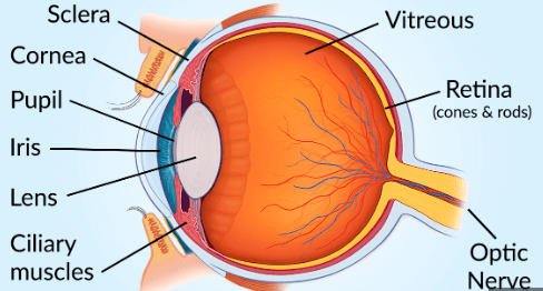
The mains parts of the Human Eye are Cornea, Iris, Pupil, Ciliary Muscles, Eye Lens, Retina, Optic Nerve, Sclera, and Vitreous Humor.
| Different Parts of the Eye | |
| Part Name | Description |
| Cornea | Transparent, clear, and dome-shaped part of the eye which helps in focusing light and maintaining the eye’s clarity. |
| Iris | A flat, colored, ring-shaped membrane of an eye that controls the size of the Pupil and regulates the amount of light entering the eye. |
| Pupil | A dark circular opening in the center of the Iris controls the amount of light entering the eye. |
| Ciliary muscles | A ring-shaped muscle that changes the shape of the lens and helps to adjust the eye’s focus for near and far versions. |
| Eye lens | A transparent, flexible structure that focuses light onto the retina, and allows us to see objects at different distances. |
| Retina | A layer of light-sensitive cells is present at the back of the eye and captures and converts light into nerve signals. |
| Optical nerve | They are a bundle of nerve fibers that carry visual information from the retina to the brain. |
| Sclera | A tough, white outer layer of the eye that provides structural support and protection to the delicate inner components of the eye. |
| Vitreous Humor | A clear, gel-like substance that fills the space between the lens and retina in the eye which helps us to maintain the shape of the eye. |
Parts of the Eye & their Functions
As we already know that the eye consists of several crucial parts. The cornea lets light in and focuses it. The iris adjusts the pupil size. The lens focuses light onto the retina, which captures the image. The optic nerve sends these images to the brain, and the other parts start their functioning. Together, these parts allow us to see and interpret the world around us. Now let us discuss all the parts of the human eye in brief
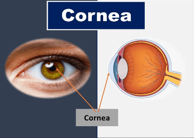
1. Cornea
The cornea is the transparent clear, dome-shaped front part of the eye. The outer surface of the cornea is convex in shape. It covers the Pupil, Iris, and anterior chamber. It is the most sensitive part of the body. The cornea plays a vital role in focusing light and maintaining the eye’s clarity. The cornea has a remarkable ability to heal quickly after minor injuries.
The cornea plays a crucial role in vision as it acts as a protective barrier and it also refracts light as it enters the eye and allows us to see clearly. The Cornea has no blood vessels and its transparency is vital for maintaining a clear vision. The curved surface of the cornea helps to focus light onto the Retina, which is very important for clear vision. Any irregularity in the shape of curvature of the cornea can lead to refractive errors like nearsightedness (Myopia), and farsightedness (Hypermetropia).
Functions of Cornea
- The cornea acts as a power convex lens, which refracts incoming light rays and helps to focus them into the retina. The retina requires this bending of light, to form a proper image.
- The cornea protects the inner structure of the eye from external elements like dust and microbes.
- The light passes through the cornea easily because of its transparency.
- The cornea also has a natural ability to absorb and filter harmful ultraviolet rays.
- The cornea does not have any blood vessels, infect it obtains nutrients and oxygen from the tear film on its outer surface.
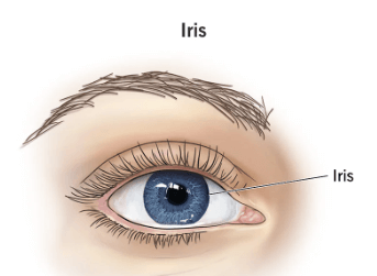
2. Iris
The Iris is present just behind the cornea. An Iris is the flat, coloured, ring-shaped membrane of an eye. The color of the Iris is determined by the amount and type of melanin pigment present in its cells, Iris gives each person their distinct eye color, such as brown, black, green, and grey.
The Iris controls the size of the Pupil and regulates the amount of light entering the eye. Like fingerprints, the pattern of the Iris is also unique to each individual. The complete study of Iris is called Iridology. There is a hole in the middle of the Iris which is called Pupil of the eye.
Functions of Iris
- The primary function of the Iris is the regulation of light that enters the eye, this takes place with the adjustment of the pupil.
- Each person has a unique eye color such as brown, black, green, and grey. The amount of pigment distributed in the iris provides color variation.
- It also acts as a physical barrier and protects the inner structure of the eye.
- The iris helps to focus incoming light into the retina.
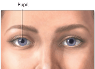
3. Pupil
The black circular opening in the center of the Iris that allow light to enter is called a Pupil. The pupil is black because no light is reflected from it. The size of the pupil changes with the amount of light, it can be explained as in bright light conditions the pupil gets smaller to reduce the amount of light that enters the eye and protects the sensitive retina from potential damage; while in low light the pupil gets larger to allow more light into the eye, which increases our ability to see the objects clearly in dim light also.
Functions of Pupil
- The Autonomic nervous system (which controls all the involuntary functions of the body) regulates the size of the pupil.
- The pupil also responds to emotional stimuli, For example: at the time of stress or anxiety, the pupil contracts; while at the time of excitement or fear, the pupil tends to expand and becomes wider.
- The size and shape of the pupil can sometimes affect the appearance of the iris color.
- Abnormal pupil responses might indicate certain neurological or ophthalmological conditions in a person.
4. Eye Lens
The eye lens is a convex lens made of a transparent, soft, and flexible material like a jelly made of protein. The eye lens is located behind the Iris. Being flexible the eye lens can also change its shape when light enters the eye through the pupil, this process is known as Accommodation. This adjustment in the shape of the lens ensures that the light rays properly focus on the retina, forming a clear sharp image.
Functions of Eye Lens
- The light rays coming into the eyes are refracted or bent by the eye lens, which helps to focus the light into the retina.
- The eye lens adjusts its curvature so that a clear sharp image is formed in the retina.
- With the help of the pupillary reflex, the eye lens adjusts its size and helps to regulate the amount of light entering the eye.
- The eye lens changes its shape to adjust the focal length, which helps us to focus on objects that are placed at a far distance. This takes place with the help of accommodation.
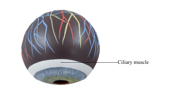
5. Ciliary Muscles
The ciliary muscle is made up of smooth muscle fibers, which means it is not under voluntary control and functions involuntarily. The ciliary muscles contract when the eye needs to focus on a nearby object, as a result, the lens becomes more round and thicker allowing it to reflect light more strongly and focus it into the retina.
The main function of the ciliary muscles is to adjust the shape of the lens. As we grow older the flexibility of the lens and the ciliary muscles decreases, which leads to Presbyopia (reduced ability to accommodate for near vision).
Functions of Ciliary Muscles
- The ciliary muscles play a huge role in the Pupillary reflex, which causes the pupil to contract when an object is placed at a very far distance.
- When looking at a distant object the ciliary muscles relax, which makes the lens flatten and decreases its refractive power.
- When looking at a near object the ciliary muscles contract, which makes the lens thicker and more curved.
- For the adjustment of the shape of the lens ciliary muscles contract and relaxes.
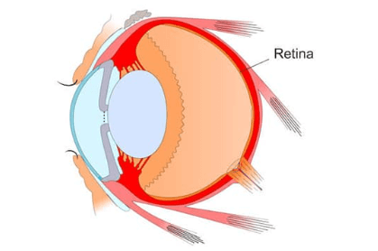
6. Retina
The screen on which the image is formed is known as Retina. The retina is present just behind the eye lens and at the back part of the eye. The retina of the eye acts the same as the film of the camera. The retina is made up of several layers of cells, each has its own specific function. The outermost layer contains specialized light-sensitive cells called photoreceptors. It is a delicate membrane and has a large number of light-sensitive cells called “Rods” and “Cones” which respond to the intensity of light and color of objects respectively by generating neural or electrical signals.
Functions of Retina
- For the detection and conversion of light retina contains specialized cells called photoreceptors (Rods and Cones).
- Visual information received from the photoreceptors is processed by the retina before sending it to the brain with the help of optic nerves.
- The cones present in the retina help us detect different colors, as cones are sensitive to different wavelengths of light.
- The Rods provide night vision which helps us to see objects clearly in low-light conditions.
- The rating contains Ganglion cells which collect processed information and send it to the brain.
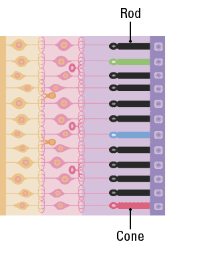
Rods
The rods refer to one of the two main types of photoreceptors. They are the rod-shaped cells present in the retina of the eye. The rods are responsible for vision in low light conditions and are light sensitive, they allow us to see the objects clearly in dim light. An Owl has a large number of rod cells in its eyes, These help the owl to see properly even in dim light during the night. Because of this, our night vision is poor as compared to an owl.
Cones
The cones are the second type of photoreceptors. They are cone-shaped cells present in the Retina of the eye. The cones are responsible for vision in bright light conditions and are sensitive to bright light, they allow us to see the objects clearly in bright light. The cone cells also respond to the colors. In other words, cone cells cause the sensation of the colors of the object in our eyes.
7. Optical Nerves
The optic nerves carry neural or electrical signals from the retina to the brain, which helps us to process visual information. Each eye has its own optic nerve, and the fibers from the left and right optic nerves partially cross over at the optic chiasm, situated near the base of the brain. This crossing over is responsible for the brain’s ability to integrate information from both eyes and create a definite visual perception.
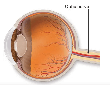
Functions of Optic Nerve
- The main function of the optic nerve is to carry visual stimuli received by the retina and send them to the brain.
- The visual cortex interrupts the signals received from the optic nerves, which helps us to recognize the shape, color, and object.
- The optic nerve fibers connect with the lateral geniculate nucleus in the thalamus and then relay the visual information.
At the junction of the Optic nerve and Retina in the eye, there are no light-sensitive cells (no rods and cones) due to which no vision is possible at that spot. This spot is known as a blind spot. Thus Blind spot is the area of the retina that is intensive to light, or we can say that a blind spot is an area in the field of vision that can’ be seen.
8. Sclera
The sclera is the tough, white, outer layer of the eye that covers most of the eye’s surface, and sclera also helps in providing structural support and protection of an eye. The sclera is primarily composed of college and elastic fibers, which makes it supportive. The primary function of the sclera is to protect the delicate inner structure of the eye, including the Retina by providing a stronger outer covering.
It also helps maintain the shape of the eyeball. The sclera is thicker at the back side of the eye (posterior) and thinnest towards the front (anterior). The sclera receives its blood supply mainly through the episcleral vessels, which are located between the sclera and the conjunctiva.
Functions of Sclera
- The sclera plays a very important role in providing structural support to the eye and it also helps in the protection of the internal parts of the eye.
- The sclera prevents the eye from collapsing under pressure.
- The sclera also acts as an attachment point for the extracellular muscles, these muscles are responsible for eye movements.
- Sclera also plays a role in focusing light on the retina by maintaining the shape of the eye.
9. Vitreous Humor
Vitreous Humor is a transparent and gelatinous substance that is filled in the space between the lens and the retina. The primary function of Vitreous Humor is to maintain the shape of the eye and also helps in the transmission of light to the retina. The vitreous humor is mostly composed of water, college fiber, and hyaluronic acid. It forms during embryonic development and remains relatively stable throughout life. However, as a person ages, the vitreous humor undergoes many changes.
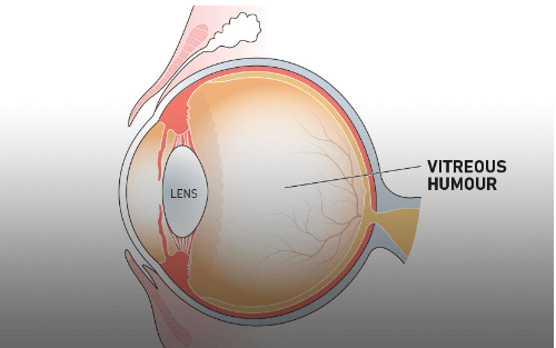
Functions of Vitreous Humor
- The vitreous Humor plays a great role in maintaining the round shape of the eye.
- Vitreous humor is transparent and allows light to pass through the eye to the retina.
- It maintains the overall refractive power of an eye, and it also helps in the transportation of nutrients and oxygen to the vascular parts of the eye.
- The vitreous humor acts as a cushion and protects all the other delicate parts of the eye.


 50 Vegetables Name for Kids in English a...
 Food Chain: Definition, Types, Examples,...
 Human Respiratory System: Definition, Di...